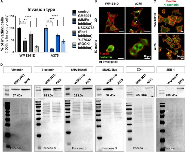
FIGURE 6.
A375 cells are more progressed in epithelial-mesenchymal transition (EMT) than WM1341D cells. (A) Analysis of 3D motility type exhibited by studied cell lines by inhibitors application: 25 μM GM6001, 10 μM NSC23766, 10 μM Y-27632. The cells were seeded onto MatrigelTM gel placed in the upper compartment of a TranswellTM in medium with added inhibitors. 24 h later the assay was terminated (n = 3). (B) Immunostainings performed on cells growing under 2D or 3D (the cells were embedded in MatrigelTM gel) conditions. F-actin and cortactin were visualized by application of fluorescently labeled phalloidin and antibodies recognizing cortactin. Arrowheads point at invadopodia. (C) Visualization of N-cadherin and F-actin in studied cells by immucytochemical staining. (D) Representative immunoblots of EMT markers levels in tested cell lines. 30 μg of protein was loaded on every lane. Membranes prior to incubation with antibodies were stained with Ponceau S to show equal protein loading on lanes. The significance level was set at ∗P < 0.05, ∗∗P < 0.01, ∗∗∗P < 0.001, and ∗∗∗∗P < 0.0001 (www.oncomine.org, February 2018, Thermo Fisher Scientific, Ann Arbor, MI, United States).

