Figure 6.
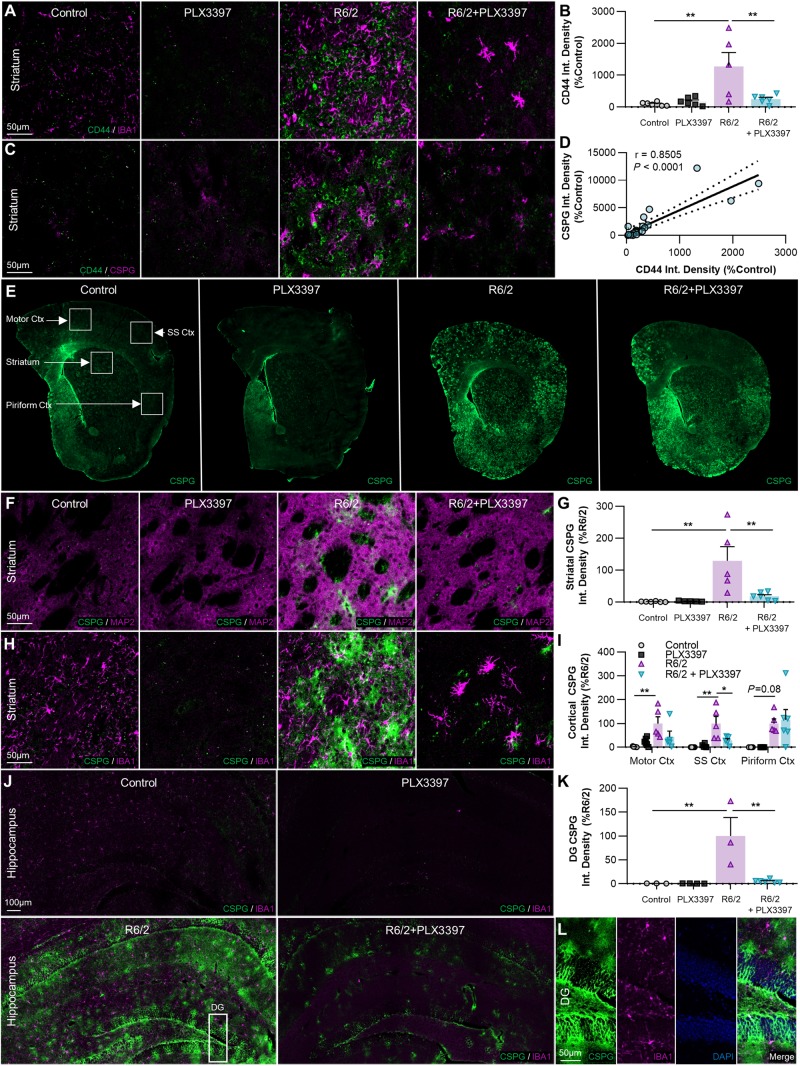
Extracellular CSPGs accumulate in the R6/2 striatum, cortex, and hippocampus, but are reduced with PLX3397. (A–D) Non-microglial expression of the cell-ECM receptor CSPG8/CD44 (×20 with IBA1+A, same images with pan-CSPG+C) is significantly increased in R6/2 striatum (B) compared to control and R6/2+PLX3397 (P < 0.01, P < 0.01) (two-way ANOVA with Tukey’s post hoc test; n = 5–6/group), and correlated with CSPG accumulation (D) across all samples (P < 0.0001, r = 0.8505) (Pearson correlation test; n = 23). (E–K) Representative whole-brain (E), ×20 striatal (with MAP2+F, same images with IBA1+H), and ×20 hippocampal stitched (J) images of CSPG+ accumulation, quantified in the striatum (G), cortex (I) and dentate gyrus (DG) of the hippocampus (K) as integrated signal density (n = 5–6/group, except dentate gyrus where n = 3–6/group). Significant increases compared to control were found in R6/2 striatum (P < 0.01), motor cortex (P < 0.01), somatosensory cortex (P < 0.01), and dentate gyrus (P < 0.01), along with a trending increase in the piriform cortex (P = 0.08) (two-way ANOVAs with Tukey’s post hoc test). Interestingly, disease-related accumulation was significantly reduced with PLX3397 in the striatum (P < 0.01), somatosensory cortex (P < 0.05), and dentate gyrus (P < 0.01) (two-way ANOVAs with Tukey’s post hoc test; n = 5–6/group, except dentate gyrus where n = 3–6/group). (L) Inset of dentate gyrus from R6/2 hippocampus (J) showing CSPG+ staining in all layers. Statistical significance is denoted by *P < 0.05, **P < 0.01. Error bars indicate SEM.

