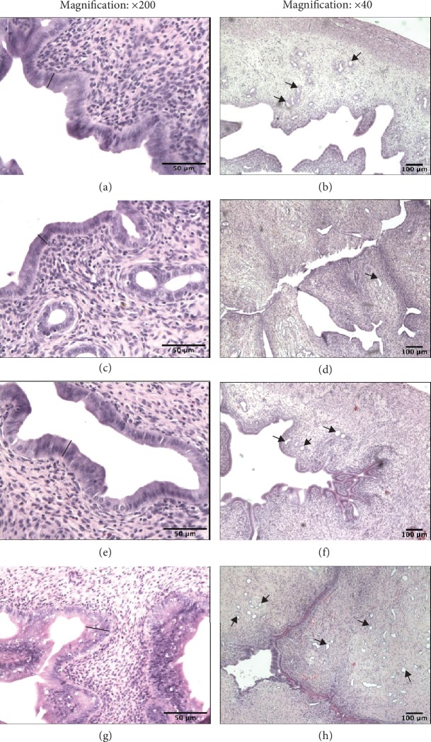
Figure 6.
Effect of CdCl2 on uterine histology. Photomicrographs of uterine sections stained with hematoxylin and eosin from rats after a 180-day postexposure period (magnification: ×200 and ×40). (a and b) Sections from the uteri of pure control rats. A thick epithelial layer is seen as well as many glands. (c) Sections from the uteri of the Cd group (4.5 mgCd/kg). The epithelial layer is thin and contains a small number of the cells. (d) Sections from the uteri of the Cd group (4.5 mgCd/kg). The epithelial layer is thin, and the glands are not numerous. (e and f) Sections from the uteri of oil control rats. The epithelial layer is similar to that of the pure control rats. (g) Sections from the uteri of the 17β-estradiol group (0.03 mgE2/kg). Notice the increased thickness of the epithelial layer. (h) Sections from the uteri of the 17β-estradiol group (0.03 mgE2/kg). Numerous uterine glands are seen. Black bars represent epithelial thickness; arrows—uterine gland.

