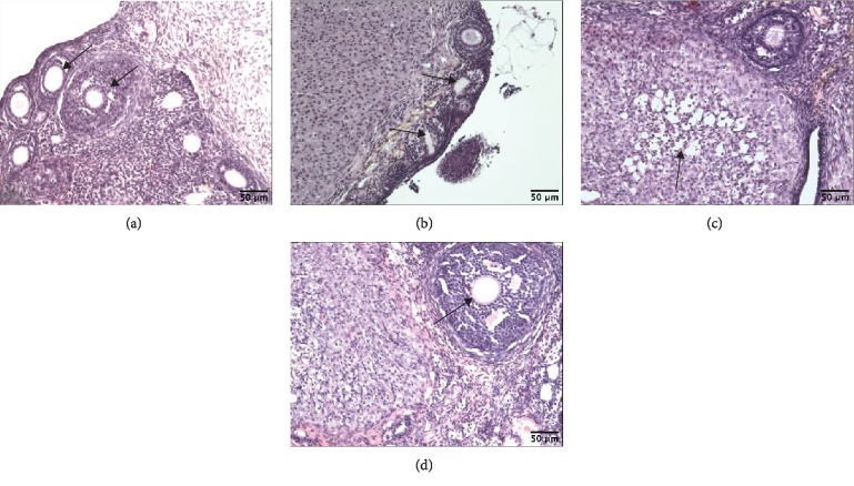
Figure 8.
Effect of CdCl2 on ovary histology. Photomicrographs of ovary sections stained with hematoxylin and eosin (magnification: ×100) from rats after a 180-day postexposure period. (a) Sections from the ovaries of pure control rats. Notice numerous oocytes (arrows). (b) Sections from the ovaries of the Cd group (4.5 mg Cd/kg). The oocytes are scanty and two of them show degenerative changes (arrows). (c) Sections from the ovaries of the Cd group (4.5 mg Cd/kg). Arrow points—degenerative changes of corpus luteum. (d) Sections from the ovaries of the 17β-estradiol group (0.03 mg E2/kg). Corpus luteum and big follicle are seen (arrow—big follicle).

