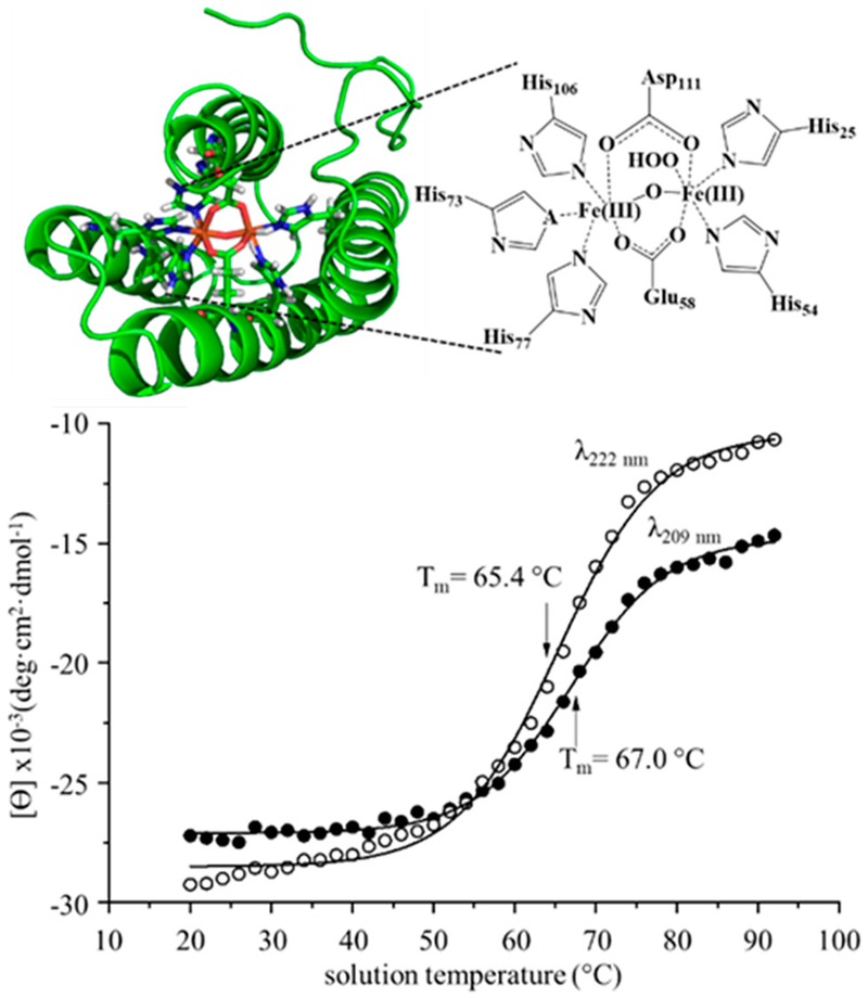
Figure 1.
Cartoon structure of the Mhr binding site (PDB structure: 2MHR) depicting the five His residues and the two bridging Asp and Glu residues involved with metal cofactor coordination (top panel). One coordination site remains available for O2 binding. Melting curves from CD data monitoring molar ellipticity (θ) at wavelengths 209 (open circles) and 220 nm (closed circles) as a function of solution temperature (bottom panel). Sigmoidal fits to the data results in midpoint melting temperatures of 65.4 ± 0.3 °C (222 nm) and 67.0 ± 0.2 °C (209 nm).

