Figure 4.

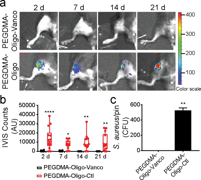
Complete eradication of S. aureus inoculated in the mouse femoral canal by PEGDMA-Oligo-Vanco coating. (a) IVIS images of mouse femurs injected with 40 CFU Xen-29 S. aureus and inserted with IM pins with PEGDMA-Oligo-Vanco or PEGDMA-Oligo coatings at 2, 7, 14, and 21 days. (b) Quantification of longitudinal bioluminescence signals of mouse femurs injected with 40 CFU Xen-29 S. aureus and inserted with the different hydrogel-coated pins at 2, 7, 14, and 21 days (n = 14). (c) S. aureus recovery from 21 day explanted pins (n = 11). Error bars represent standard deviations. * p ≤ 0.05, ** p ≤ 0.01, *** p ≤ 0.001, **** p ≤ 0.0001 (two-way ANOVA for part b; Student’s t-test for part c).
