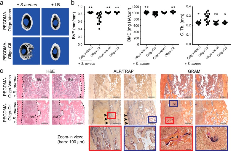
Figure 5.
Prevention of the development of osteomyelitis in mouse femoral canal inoculated with S. aureus by PEGDMA-Oligo-Vanco coating. (a) 3D μCT axial images of the distal femoral region 21 days after the insertion of Ti6Al4V IM pins (pins excluded during contouring) with different hydrogel coatings, with or without the inoculation of 40-CFU Xen-29 S. aureus. (b) Quantification of femoral BVF, BMD, and C. Th. of infected and uninfected femurs 21 days after the insertion of Ti6Al4V IM pins with PEGDMA-Oligo-Vanco or PEGDMA-Oligo coatings. n = 11–14. Error bars represent standard deviations. * p ≤ 0.05, ** p ≤ 0.01 as compared to the PEGDMA-Oligo control coating + S. aureus group (one-way ANOVA). (c) H&E, ALP (blue)/TRAP (red), and Gram staining (bacteria stain blue) of explanted femurs in the infected group with PEGDMA-Oligo-Vanco coating or PEGDMA-Oligo control coating at 21 days postoperation. Dashed lines outline the cortical bone; BM = bone marrow; BM* = infected bone marrow; arrowheads indicate regions of enhanced ALP/TRAP activities; higher magnification views of the regions within the blue and red boxes are shown in the bottom row. Scale bars = 500 μm (top and middle rows) or 100 μm (bottom row).

