Figure 1.
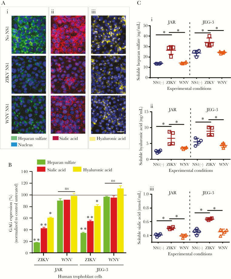
Zika virus (ZIKV) NS1 induces shedding of gycosaminoglycans (GAGs) from human trophoblast cells. (A) Immunofluorescence staining of (i) heparan sulfate [HS], (ii) sialic acid [Sia], and (iii) hyaluronic acid [HA] in confluent monolayers of JAR cells cultured on collagen-treated glass cover slips after 24 hours of treatment with ZIKV NS1 Suriname (5 µg/mL) or West Nile virus (WNV) NS1 (5 µg/mL). Images are representative of 4 independent experiments run in duplicate. Magnification, x20. Scale bar = 10 µM. (B) Mean fluorescence Intensity analyses of GAG expression on JAR monolayers after ZIKV or WNV NS1 treatment as described above. Each bar shows the mean ± standard error (SE) of 4 independent experiments processed in duplicate. The percentage of GAG expression on JAR and JEG cells treated with NS1 was normalized to the control untreated cells taken as 100%. (C) Enzyme linked immunosorbent assay quantification of soluble (i) HS, (ii) HA, and (iii) Sia in the supernatants obtained from JAR and JEG-3 cells 24 hours posttreatement with ZIKV (open squares) or WNV NS1 (open triangles) proteins (5 µg/mL). Untreated cells (open circles). Data were derived from 4 independent experiments and were analyzed by nonparametric Mann-Whitney analysis. *, P < .05. Magnification, x20. Scale bar = 10 µM. ns, not significant.

