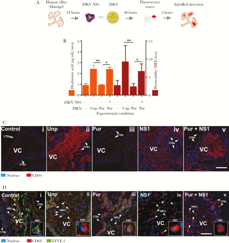
Figure 4.
Zika virus (ZIKV) in the presence of NS1 induces explant permeability, hyaluronic acid (HA) degradation, and CD44 upregulation in chorionic villous explants. (A and B) Permeability to Alexa680-dextran and levels of HA were measured in chorionic placental explants (n = 3) exposed to either unpurified ZIKV (Unp) or purified ZIKV (Pur) in the presence or absence of ZIKV NS1 (5 µg/mL) at 2 days postinfection. Raw mean fluorescence intensity (MFI) values were normalized to the cross-sectional area of the explant (MFI/area) (right y-axis). For permeability values, levels of HA (left y-axis) were plotted side-by-side with area-normalized permeability values (right vertical scale). (C and D) Side-by-side immunofluorescent staining of CD44 and lymphatic endothelial cell HA receptor (LYVE)-1 in sections of chorionic villous explants treated and infected as in A and B. Data shown are the MFI/area values for each explant normalized to each explant size, including the mean ± standard error. CD68 was used as a marker for fetal macrophages, Hofbauer cells ([HBC] red) (D). Zoomed-in inset depicting expression of CD68 and LYVE-1 in HBCs (lower right). Explants treated only with ZIKV NS1 were also included (iv). Images are representative of 2 independent experiments. Magnification, ×40. Scale bar = 100 µM.

