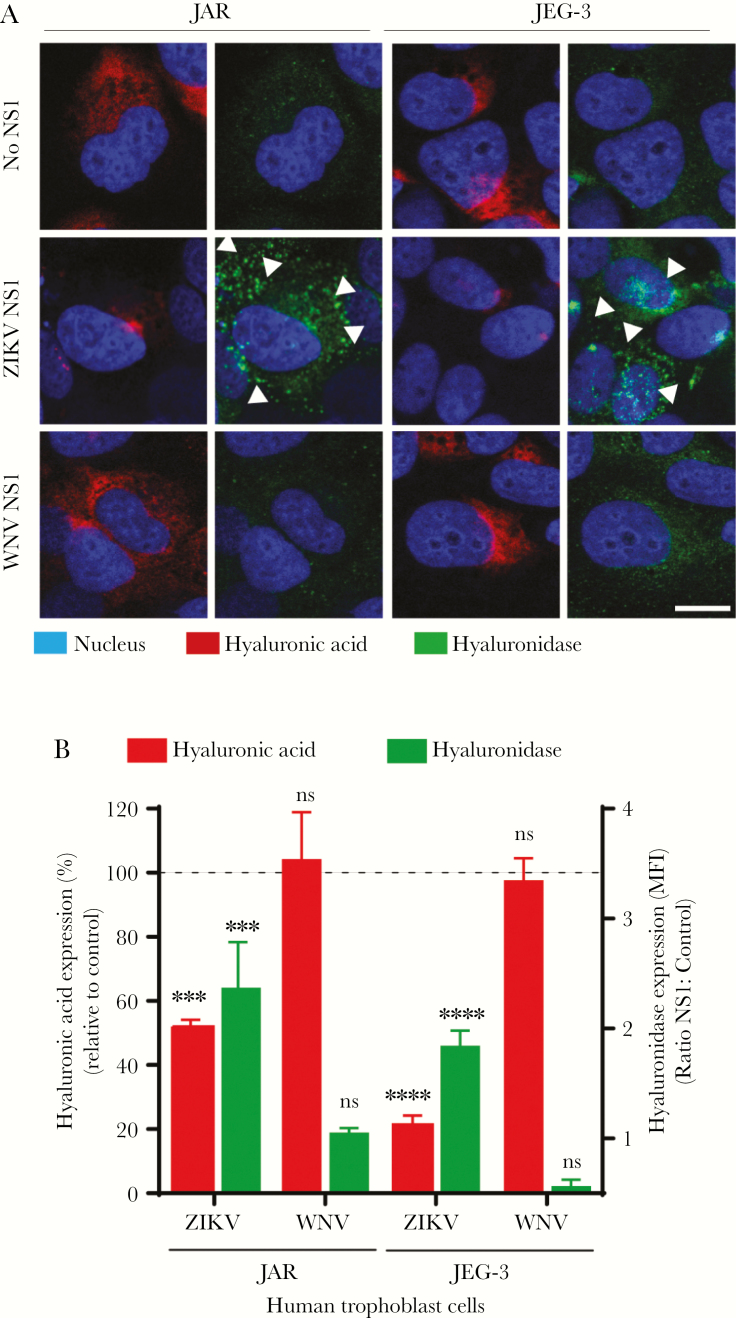
Figure 5.
Zika virus (ZIKV) NS1 modulates the expression of hyaluronidases in human trophoblast cells. (A) Immunofluorescence staining of hyaluronic acid ([HA], first and third columns) and human hyaluronidase ([HYAL-1], second and fourth columns) in confluent monolayers of JAR and JEG-3 cells cultured on collagen-treated glass coverslips 24 hours posttreatment with ZIKV NS1 (5 µg/mL) and West Nile virus (WNV) NS1 (5 µg/mL). Images are representative of 2 independent experiments run in duplicate. Magnification, x20. Scale bar = 10 µM. White arrowheads indicate expression of HYAL-1. (B) Mean fluorescence intensity analyses of HA and hyaluronidase expression in JAR and JEG-3 cultures after ZIKV or WNV NS1 treatment as described above. Each bar shows the mean ± SE of 2 independent experiments run in duplicate. The percentage of HA expression (dark gray) in JAR and JEG-3 cells treated with NS1 was normalized to the control untreated cells taken as 100%. Hyaluronidase (light gray) was expressed as the ratio (fold change) between NS1 and untreated cells used as control. White arrowheads indicate hyaluronidase expression puncta in trophoblast cells (light gray). Nuclei were stained with Hoechst. Magnification, x20. Scale bars= 5 µM. Ordinary one-way analysis of variance was used for statistical analyses: ***, P < .001; t test, ****, P < .0001 (ZIKV NS1 vs control: JAR and JEG-3 cells). ns, not significant.

