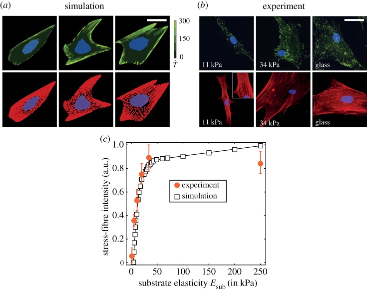
Figure 2.
(a) Predictions from simulations and (b) observations from Engler et al. [6] of hMSCs seeded on elastic substrates uniformly coated with collagen. In the experimental immunofluorescence images, the focal adhesions are coloured green, actin red and nucleus blue and purple, and a similar scheme is followed in the predictions, with the focal adhesions parametrized by the magnitude of the normalized traction (see electronic supplementary material, s1.5.1 for details of the method used to construct immunofluorescence-like images from the simulated results). Scale bar, 20 µm. (c) Predictions of stress-fibre intensity as a function of substrate stiffness compared with observations from Zemel et al. [36] for hMSCs seeded on glass substrates. In the simulations, we use total stress-fibre concentration to parametrize the stress-fibre intensity. (Online version in colour.)

