Figure 7.
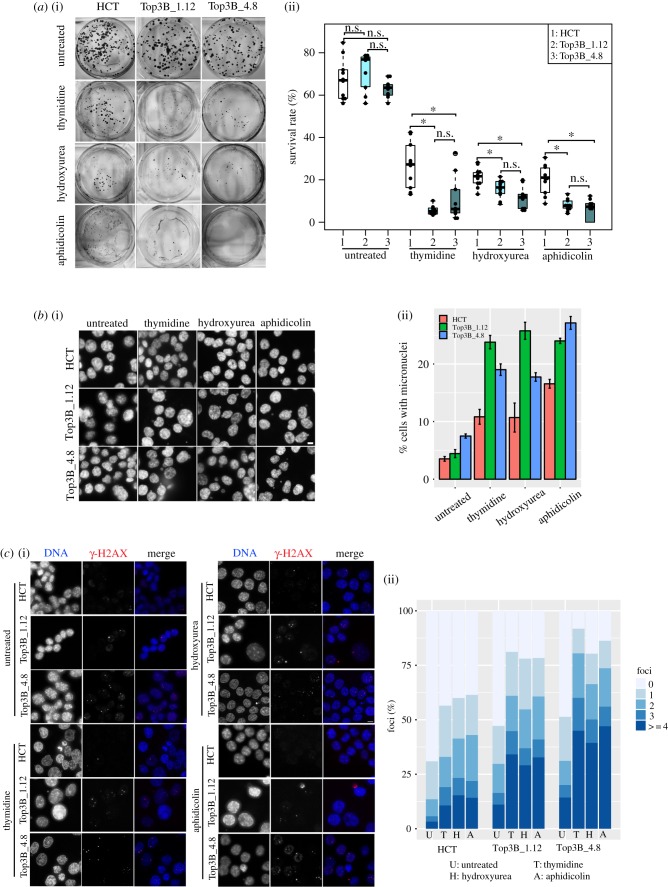
Recovery after replication stress is impaired in TOP3B null cells. (a) (i) Representative images of crystal violet-stained plates from the colony-forming assay. HCT116 control (HCT) and HCT116 TOP3B null (Top3B_1.12 and Top3B_4.8) cells were treated with no drug, thymidine, hydroxyurea or aphidicolin and allowed to recover for seven days. (ii) Survival rate (%) of colony formation. Data are from three independent experiments, with each experimental sample done in triplicate or more. Asterisk denotes p < 0.05. n.s., no significant difference. (b) (i) Representative DNA-stained (DAPI) images of micronuclei formation of HCT116 control and HCT116 TOP3B null cells treated with no drug, thymidine, hydroxyurea or aphidicolin and allowed to recover for 48 h. (ii) Quantification of micronuclei. Data are from three independent experiments, with at least 1000 cells scored at each experiment. (c) (i) Representative images of HCT116 control and HCT116 TOP3B null clones treated with no drug, thymidine, hydroxyurea or aphidicolin and allowed to recover for 48 h and stained for γ-H2AX (red) and DNA (DAPI, blue). (ii) Quantification of the number of γ-H2AX foci in each cell. Data are from three independent experiments, with at least 150 cells scored at each experiment. Scale bar, 5 µm.

