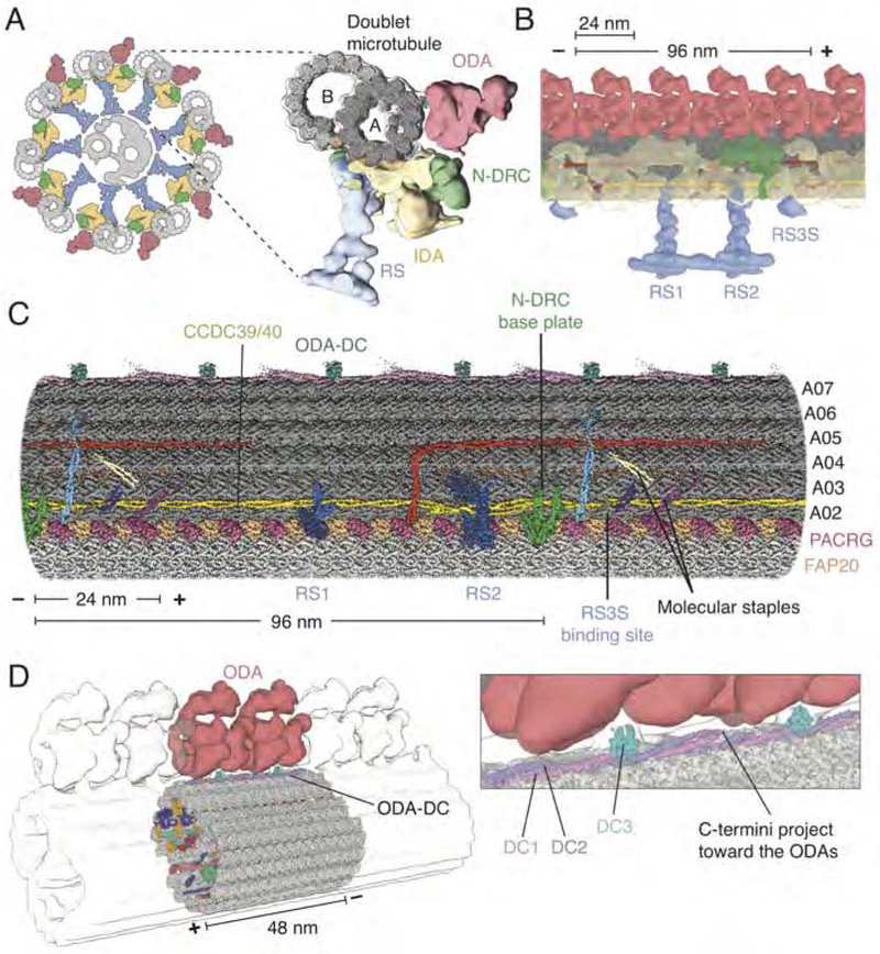
Figure 1. Structure of the 96-nm Repeat of the Doublet Microtubule and Relationship to the Axoneme.
(A) Left, schematic representation of the cross-section of the axoneme from C. reinhardtii showing nine doublet microtubules surrounding a central pair of singlet microtubules (grey). Attached to the doublet microtubules are the radial spokes (RS; blue), inner dynein arm (IDA; yellow), nexin-dynein regulatory complex (N-DRC; green) and outer dynein arm (ODA; red). Right, subtomogram average (EMD-6872) (Kubo et al., 2018) of the axoneme with our map of the 96-nm doublet microtubule repeat (grey) docked inside.
(B) Longitudinal view of the doublet microtubule docked into the subtomogram average of the axoneme (EMD-6872).
(C) Density map of the 96-nm repeat showing axonemal proteins decorating the external surface of the A tubule.
(D) The ODA-DC repeats every 24 nm and, based on fitting the structure into the subtomogram average (EMD-6872), is the main attachment point for the ODA. Inset, detail of the interaction between the ODA and ODA-DC.
In all panels, the minus (−) and plus (+) ends of the doublet microtubule are indicated at the ends of the scale bar.
See also Figures S1, S2, S3, S4 and Tables S1, S2, S3 and S4.

