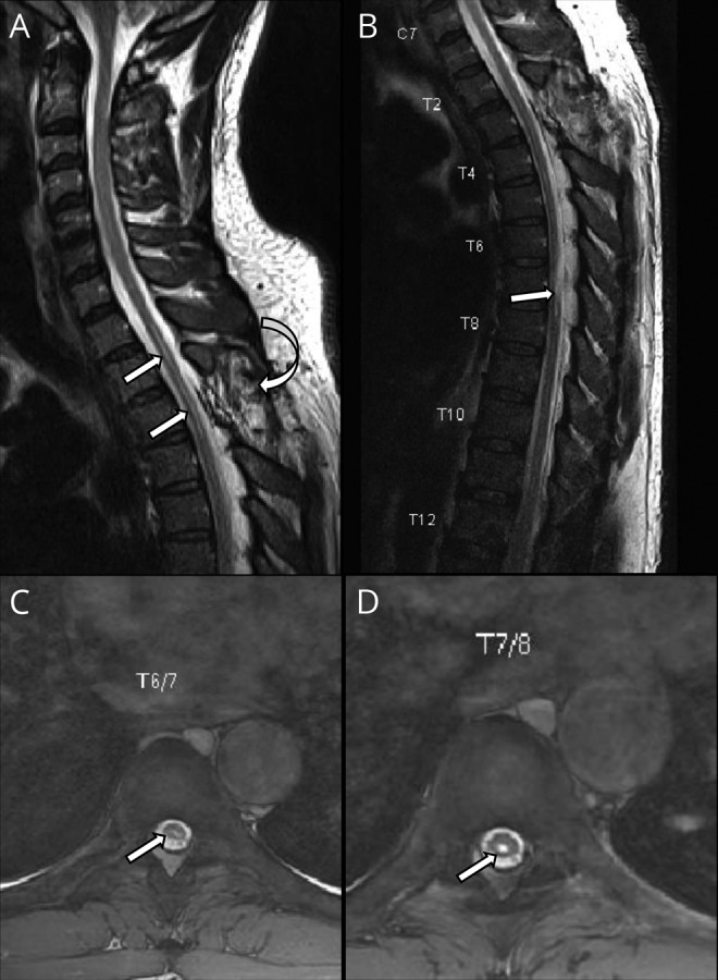
Figure 1. Patient's dorsal spine MRI showing cord findings and previous intervention.

(A) Sagittal T2WI of the dorsal spine showing the site of the previous surgery (curved arrow) and intramedullary syrinx of the upper thoracic cord (arrows). (B) Sagittal and (C and D) axial T2WI of the thoracic spine showing cord affection by intramedullary patch of hyper intensity (arrows).
