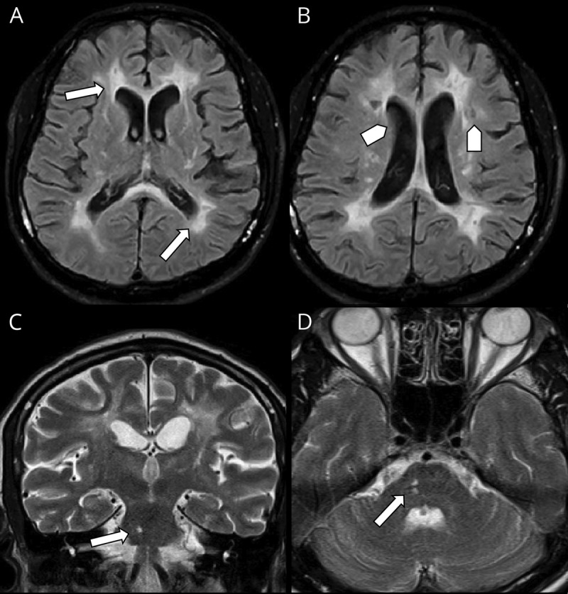
Figure 2. Patient's brain MRI showing hemispheric and brainstem findings.

Pattern of periventricular white matter involvement in different pulse sequences in axial FLAIR (A and B), note lacunar infarcts (arrow head) is seen in B. (C) Coronal T2WI and (D) Axial T2WI showing affection of the right aspect of pons (arrows) creeping to the middle cerebral peduncle. FLAIR = fluid-attenuated inversion recovery.
