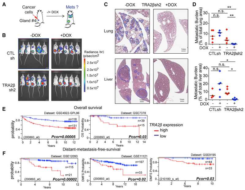
Figure 7. TRA2β Plays a Role in Metastasis In Vivo in a TNBC Mouse Model and in Breast Cancer Patients.
(A) CTLsh or TRA2βsh2MDA-MB231 cells are injected into the mammary fat pad of NSG mice; primary tumors and metastasis are monitored by bioluminescence imaging.
(B) Bioluminescence detection of primary tumors and metastasis in mice injected with CTLsh or TRA2βsh2 MDA-MB231 cells ± DOX at 8 weeks post-injection.
(C) Representative H&E lung and liver sections of mice injected with TRA2βsh2 MDA-MB231 cells ± DOX (scale bar: 1 mm). Metastatic areas are circled in blue.
(D) Quantification of metastasis burden in mice injected with CTLsh or TRA2βsh2 MDA-MB231 cells ± DOX (n ≥ 4; t test, *p < 0.01, **p < 0.001, n.s., not significant).
(E and F) Correlation between TRA2β expression and overall survival (E) or distant metastasis-free survival (F) in cohorts of breast cancer patients stratified by TRA2β levels. Cohort size, dataset, and probe ID, and corrected p value (log-rank test) are indicated.
See also Figure S7.

