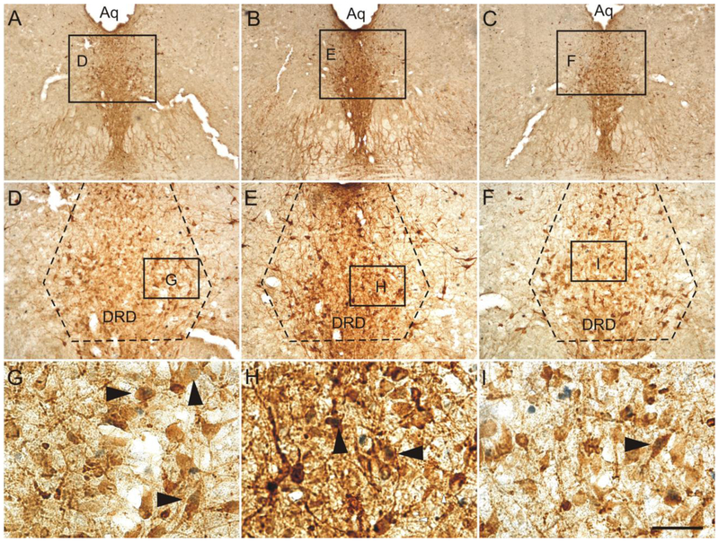
Fig. 9.
Photomicrographs illustrate c-Fos (PC38)/tryptophan hydroxylase (Tph) immunostaining in the dorsal raphe nucleus (−7.64 mm bregma) in representative rats from each treatment group in Experiment 2. Photomicrographs illustrate immunostaining in rats exposed to (A, D, and G) sedentary conditions, (B, E, H) voluntary exercise, and (C, F, and I) forced exercise. Black boxes in A, B, and C indicate regions displayed at higher magnification in D, E, and F. Black boxes in D, E, and F indicate regions displayed at higher magnification in G, H, and I. Black arrowheads indicate c-Fos (PC38)-immunoreactive/Tph-immunoreactive neurons (brown cytoplasmic staining with blue/black nuclear staining). Abbreviations: DRD; dorsal raphe nucleus, dorsal part; Aq; cerebral aqueduct. Scale bar: A-C, 500 μm; D-F, 100 μm; G-I, 25 μm.

