Fig. 6.
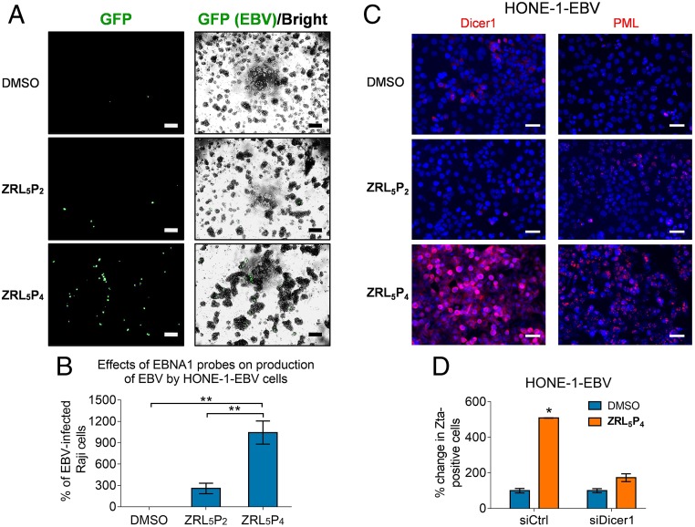
Production of infectious EBV particle in response to ZRL5P4. The HONE-1-EBV cell line, which expresses GFP to indicate the presence of the EBV genome, was used. This cell line was treated with 10 µM ZRL5P2 or ZRL5P4 for 4 d, and the viral particles released in the culture medium were detected by the Raji cell assay. The culture medium was added to Raji cells for 3 d, and GFP expression reflects the reinfection by the HONE-1–released EBV particles. (A) Representative results are shown. The GFP signal was detected by ultraviolet light exposure, and the cell morphology was captured by phase-contrast light microscopy and the bright-field image was merged with the GFP image. Magnification, 400×. (Scale bars, 100 µm.) (B) Relative average viral titer in response to ZRL5P2 or ZRL5P4 was compared with the solvent control (DMSO). The Raji cell assay was performed in triplicate for each treatment. **P < 0.01, statistically significant difference. Data are expressed as the means ± SD. (C) Representative images of immunofluorescent analysis of Dicer1 and PML in HONE-1-EBV cells in response to 10 µM ZRL5P2 or ZRL5P4. The nuclei were counterstained with DAPI and indicated in blue. (Scale bars, 50 µm.) (D) Comparison of the number of Zta-positive cells after treatment with ZRL5P4 in the presence versus the absence of Dicer1. HONE-1-EBV and NPC43 were included. Gene silencing of Dicer1 was achieved by siRNA transfection. Zta was detected by immunofluorescent analysis. Control siRNA (siCTL) was used as a negative control. Relative percentage of Zta-positive was compared against the DMSO solvent control in the siRNA control cells. *P < 0.05.

