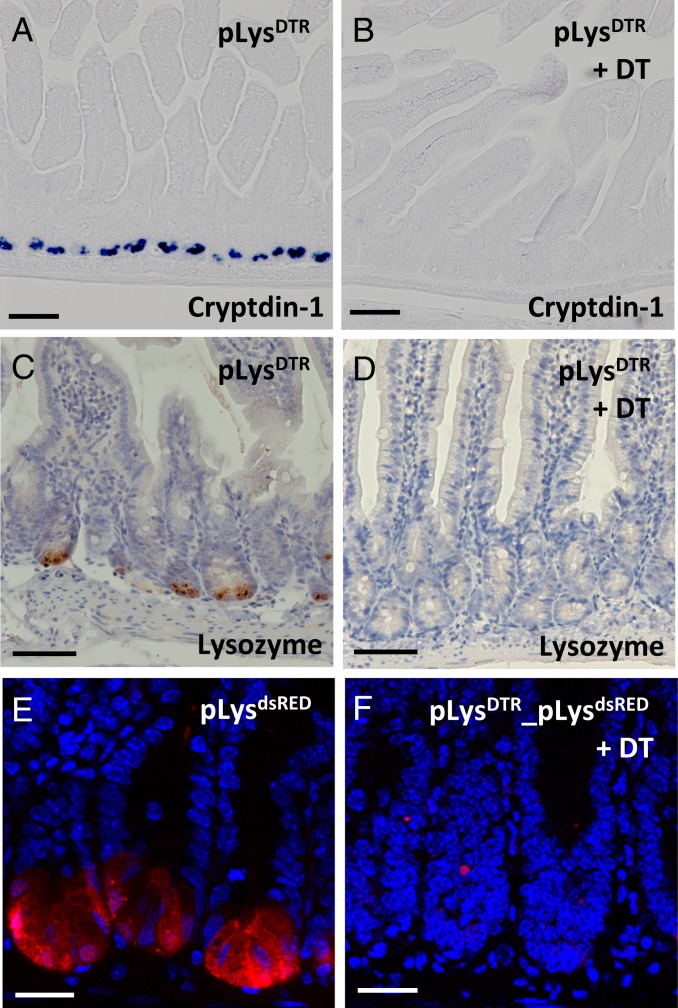
Fig. 2.
Efficient deletion of Paneth cells in the pLysDTR KI mice on DT administration. In situ hybridization of cryptdin-1 (A and B), lysozyme-specific immunostaining (C and D), and confocal imaging for dsRED expression (E and F) on jejunum sections from the untreated pLysDTR KI (A and C) and pLysdsRED (E) control mice and the pLysDTR KI (B and D) and pLysDTR_ pLysdsRED (F) mice DT-treated for 6 consecutive days, at 16 h after the last DT injection. The analysis shows the absence of Paneth cells in the intestines of DT-treated pLysDTR mice (B and D) and pLysDTR_pLysdsRED mice (F), in contrast to the controls (A, C, and E). (Scale bars: 100 µm in A–D; 50 µm in E and F.)

