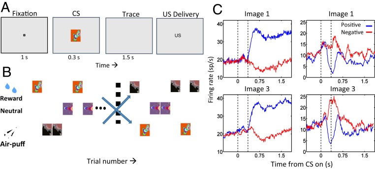
Fig. 1.
Amygdala neurons represent the positive and negative valence of CSs. (A) Sequence of a trace-conditioning trial. Monkeys centered gaze at a fixation point for 1 s and viewed a fractal image for 300 ms. US delivery followed a 1,500-ms trace interval. (B) Task structure. Positive images, liquid reward; negative images, aversive air puff; nonreinforced images, no US. After monkeys learned initial contingencies, image reinforcement contingencies switched without warning. (C) PSTHs for 2 neurons. Reward trials, blue; air puff trials, red. Image 1 was initially rewarded, then paired with air puff after reversal; image 3, opposite contingencies. (Left column) Positive value-coding neuron, responding more strongly to both images when rewarded. (Right column) Negative value-coding neuron, responding most strongly when air puff follows each image presentation. Reprinted with permission from ref. 28.

