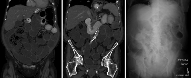
Figure 2.

(A,B) Coronal reformations of an abdominopelvic CT show that the small bowel is fluid filled from duodenum through the ileocecal valve with many loops mildly dilated. Oral contrast that had begun being administered 1 hour prior to imaging had not progressed beyond the proximal jejunum. (C) Supine abdominal radiograph 1 month subsequent shows similarly mildly dilated loops of small bowel with air and fluid with stool having passed from the colon, which is now distended with gas in portions.
