Fig. 1.
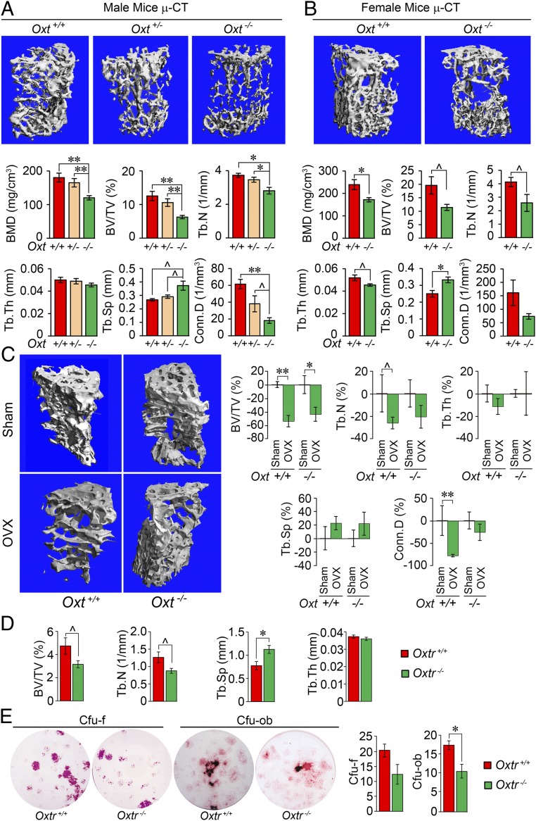
OXT deficiency reduces bone mass but does not affect ovariectomy-induced bone loss. Representative µCT images and trabecular bone microstructural parameters—BMD, BV/TV, Tb.N, Tb.Th, trabecular spacing (Tb.Sp), and Conn.D—of 10-mo-old male (A) and female (B) wild-type (+/+), heterozygotes (+/−), or homozygotes (−/−) for the oxytocin gene, Oxt (n = 4 to 8 mice per group). (C) Representative µCT images and microstructural parameters (% control) showing the effect of ovariectomy (OVX) or sham operation (Sham; control) on 2-mo-old Oxt+/+ and Oxt−/− mice (n = 3 to 9 per group). (D) µCT-based trabecular bone microstructural parameters in female wild-type and Oxtr-deficient (Oxtr−/−) mice (n = 3 to 4 per group). (E) Representative cultures and quantitation of CFU-F stained for alkaline phosphatase and CFU-OB labeled with von Kossa stain. The colonies formed when bone marrow stromal cells isolated from Oxtr+/+ or Oxtr−/− mice were allowed to grow in differentiation media (β-glycerol phosphate, ascorbic acid, and dexamethasone) for 7 and 21 d, respectively. Colonies per well were counted in triplicate. Data are expressed as mean ± SEM; comparisons with control mice, or as shown; *P < 0.05, **P < 0.01, or showing a trend ^0.05 < P < 0.1, 2-tailed Student’s t test or one-way ANOVA with Holm–Sidak correction.

