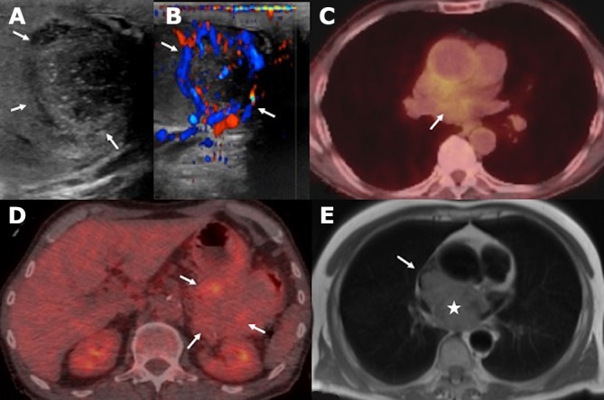
Figure 2.

(A, B) Testicular ultrasound with B-mode and colour Doppler shows a heterogeneous, hyporeflective lesion with peripheral vascularity (arrows). (C, D) PET/CT images show moderate FDG uptake of the mediastinal mass and mild metabolic activity of the gastric mass (arrows). (E) Axial post-gadolinium T1 MR imaging of the thorax confirms the aggressive features of the mediastinal mass with right atrial (asterisk) and superior vena cava invasion (arrow).
