Fig. 4.
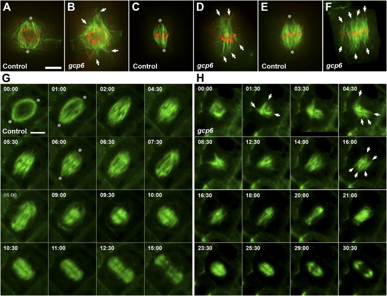
GCP6 plays a critical role in spindle morphogenesis. (A–F) Comparative views of MT organization in wild-type control cells (A, C, and E) and gcp6 mutant cells (B, D, and F) at late prophase (A and B), prometaphase (C and D), and metaphase (E and F). Merged micrographs have MTs in green and DNA in red. (A) MTs formed on the nuclear envelope are organized into a prospindle centered at two poles (asterisks) in a control cell when the preprophase band MTs are barely detected. (B) In the gcp6-2 mutant cell at a similar stage, MT bundles are disorganized and aligned toward discrete foci (arrows). (C) After the nuclear envelope breakdown, MT bundles start to encounter condensed chromosomes and assume the spindle array with convergent poles (asterisks) in the control cell. (D) The gcp6-1 mutant cell also forms MT bundles that have contacts with chromosomes but point at different directions (arrows). (E) When chromosomes are aligned at the metaphase plate, a typical acentrosomal spindle MT array has convergent poles (asterisks). (F) The gcp6-1 mutant cell forms a spindle array with nearly parallel MT bundles (arrows) connected to chromosomes aligned at the metaphase plate. (G and H) Comparative views of cell cycle-dependent MT reorganization in control (G, from Movie S2) and a gcp6 mutant cell (H, from Movie S3) that express the VisGreen-TUB6 fusion protein. The time stamps are in min:s. (G) In the control cell, spindle poles can be discerned (asterisks). At prophase, the prospindle exhibits a clear bipolar configuration on the nuclear envelope (00:00), followed by rigorous MT formation and bundling after the nuclear envelope breakdown (01:00 to 04:30). At 05:30, a clear metaphase spindle is established, followed by shortening of kinetochore fibers at 06:00 to 08:00. In the meantime, rich MTs are formed in the spindle midzone (06:00 to 09:00). These MTs eventually assume a mirrored pattern leaving a dark midline, marking the birth of the phragmoplast MT array (09:00). This phragmoplast array expands toward the cell periphery while becoming shortened (09:30 to 15:00). Eventually, MTs begin to be disassembled from the center (15:00). (H) In the gcp6-1 mutant cell, MTs are formed on the nuclear envelope but not in an obvious prospindle configuration (00:00). Rigorous MT polymerization takes place, resulting in the formation of randomly oriented MT bundles (arrows) following nuclear envelope breakdown (01:00 to 12:30). Eventually, a bipolar array can be seen but without MTs converging toward the poles (14:00). A metaphase spindle is established with more or less parallel kinetochore fibers (arrows; 16:00). At anaphase, the shortening of kinetochore fibers is concomitant with the formation of MTs in the spindle midzone (18:00 to 21:00). These spindle midzone MTs are replaced by a mirrored phragmoplast array with a dark line in the middle (23:30). This cytokinetic array expands while having its MTs shortened and depolymerized from the center, as seen in the control cell (25:30 to 30:30). (Scale bar: 5 µm.)

