Fig. 3.
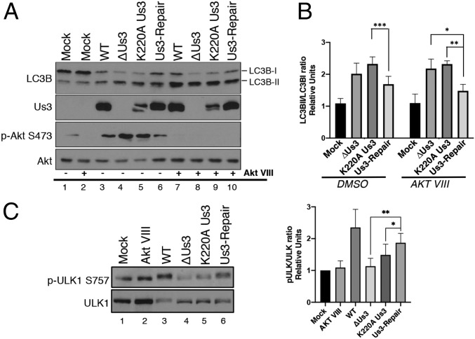
Us3 acts downstream of ULK1. (A) NHDFs were mock infected or infected (MOI = 5) with wild-type HSV-1 (WT), a Us3-deficient virus (ΔUs3), a virus expressing a catalytically inactive Us3 protein (K220A Us3), or a virus where the Us3 deficiency was repaired (Us3-Repair). After 1.5 h cells were treated with an Akt inhibitor (Akt VIII) or DMSO. At 12 hpi, total protein was fractionated by SDS/PAGE and analyzed by immunoblotting with the indicated antibodies. Migration of unmodified LC3B-I and lipidated LC3B-II are indicated on the Right of this representative image. (B) The LC3BII/LC3BI ratio from 4 (n = 4) separate experiments (A) was quantified using ImageJ and plotted. Error bars = SEM. *P < 0.05; **P < 0.01; ***P < 0.001 by 2-way ANOVA corrected for multiple comparisons. (C) Phosphorylation of endogenous ULK1 in NHDFs infected as in A (Left shows representative image) and the ratio of phosphorylated p-ULK1 S575 to total ULK1 quantified from 5 separate experiments (n = 5) using Licor Image Studio software (Right). Error bars = SEM. *P < 0.05; **P < 0.005 by 2-way ANOVA corrected for multiple comparisons.

