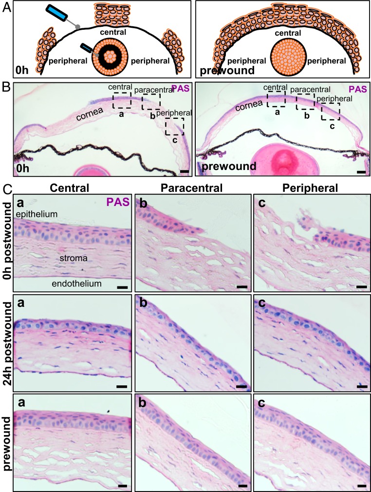
Fig. 1.
Corneal epithelial restoration after an annular debridement injury. (A) Schematic depiction on how the annular epithelial wound was inflicted. (B) WT mice (n = 4/group/time-point) had their right corneal epithelium mechanically debrided to inflict a type I annular defect. Representative images of transverse sections stained with PAS provide a panoramic overview of tissue architecture immediately after (Left) and prior to (Right) wounding. (Scale bars, 200 μm.) (C) The region encompassed by the hatched squares in B is magnified in (a) central, (b) paracentral, and (c) peripheral zones, respectively. Displayed are representative images taken immediately after wounding (first row), 24 h postwounding (second row), and prior to injury (third row). (Scale bars, 20 μm.)

