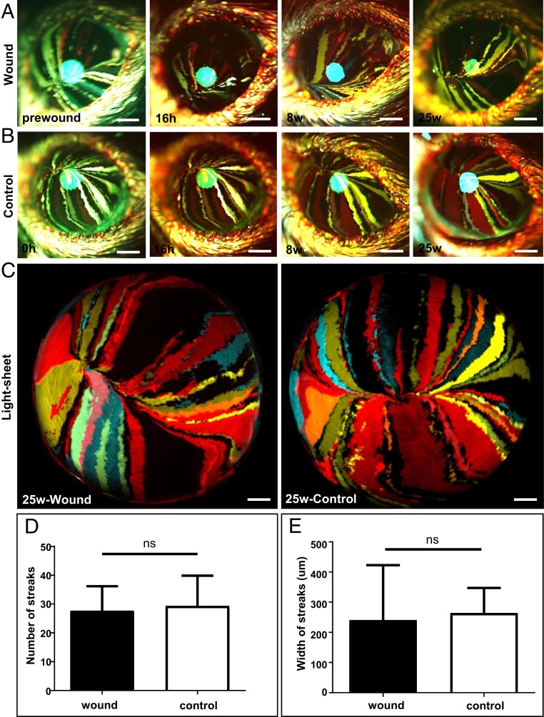
Fig. 3.
Cells involved in the long-term maintenance of corneal epithelium after wound closure. (A) Confetti mice at 30-wk posttransgene induction (n = 3/group) had their right corneal epithelium mechanically debrided to create a type II annular wound, after which they were monitored long-term. Corneas were imaged by intravital microscopy before and at 16 h, 8-wk, and 25-wk postinjury. (Scale bars, 400 μm.) (B) The intact left eye was used as the control. (Scale bars, 400 μm.) (C) A further set of corneas (n = 3/group) were procured from euthanized mice and imaged by light-sheet microscopy 25-wk after inflicting a type II annular injury. (Scale bars, 500 μm.) (D and E) Streak number and width (respectively) at 25-wk postinjury (mean ± SD, n = 3/group, ns = not significant; P > 0.05, unpaired 2-tailed Welch’s t test).

