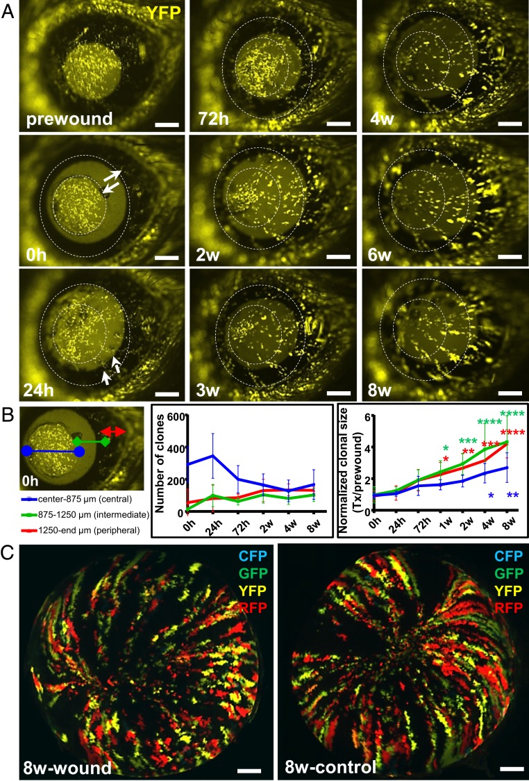
Fig. 4.
Reepithelialization of annular wounds by centripetally migrating K14+ limbal-derived clones. (A) One week after transgene induction, Confetti mice (n = 5/group) had their right corneal epithelium mechanically debrided to create a type I annular wound. For ease of visualization, only YFP+ (yellow) luminescing cells were monitored long-term by intravital microscopy. Solid white arrows indicate the wound span immediately (0 h) after injury. Small white arrows point to peripheral YFP+ clones moving centripetally across the wound bed 24 h postinjury. Hatched white circles indicate the wounded region. (Scale bars, 400 μm.) (B) YFP+ patches and clones located in the central (apex-to-875 μm, blue), intermediate (875 μm-to-1250 μm, green), and peripheral (1250 μm-to-eyelid margin, red) zones were analyzed, and their number and size displayed. Data represent mean ± SD, n = 5/group, *P < 0.05, **P < 0.01, ***P < 0.001, ****P < 0.0001, 2-way ANOVA with Dunnett’s multiple comparisons test. (C) After enucleation, the same corneas and their contralateral control counterparts were imaged by light-sheet microscopy at 8-wk postinjury. (Scale bars, 500 μm.)

