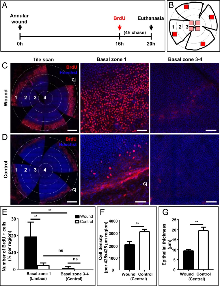
Fig. 5.
Proliferation of peripheral and central corneal epithelia after an annular injury. (A) Schematic diagram shows the experimental design whereby WT mice (n = 4/group) had their right corneal epithelium debrided to generate a type II annular wound. After 16 h, mice were injected with BrdU, euthanized 4 h later, then corneas were procured and flat-mounted. (B) Schematic diagram shows a flattened cornea in which 4 equally distant concentric rings are drawn, and zones numbered accordingly; 1 (limbus), 2 (peripheral), 3 (paracentral), and 4 (central). BrdU+ basal epithelia within zone 1 and zones 3 and 4 were counted in each of 4 randomly selected regions that measured 425 × 425 μm in area (red squares). (C and D) Representative confocal images of wounded and control corneas stained for BrdU (red) and counterstained with Hoechst (blue). (Scale bars, 500 μm [tile scan] and 100 μm [basal layer of zones 1, and 3, and 4].) (E–G) Bars in the graphs represent percentage BrdU+ basal epithelia in zone 1 (limbal) compared to zones 3 and 4 (paracentral and central), and basal cell density and epithelial thickness within the intact central epithelial island comparing wounded to controls (mean ± SD; n = 4/group independent corneas, **P < 0.01, 2-way ANOVA with Tukey’s multiple comparisons; ns = not significant).

