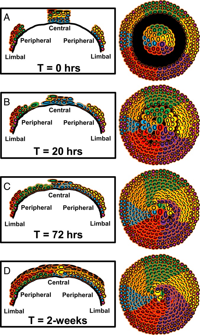
Fig. 7.
Schematic representation of how annular epithelial wounds heal. The model is based on results obtained from Confetti mouse corneas where K14+ LESC-derived clones are distinguishable as multicolored colonies that form curvilinear stripes. The first column is a cross-sectional view of the cornea spanning limbus-to-limbus. The second column provides an overview of the entire cornea from an apical perspective. Each row (A–D) corresponds to a specific time point after injury. (A) Time (T) = 0 h; represents the time when corneas are debrided to inflict an annular wound (black region). (B) T = 20 h; proliferation in the limbus increases (pink nuclei) and cells move into the wound bed from the periphery. Cells at the advancing edge are flatter and larger. Cells on the fringe of the central island do not proliferate or move, rather they change shape due to the extra space available. This shape change coincides with a thinner corneal epithelium within the central island. (C) T = 72 h; proliferation subsides, and epithelial cells have filled in the void. They begin stratifying and replacing cells from the central island. (D) T = 2-wk; epithelial layering is restored and only a few of the original central epithelial cells remain.

