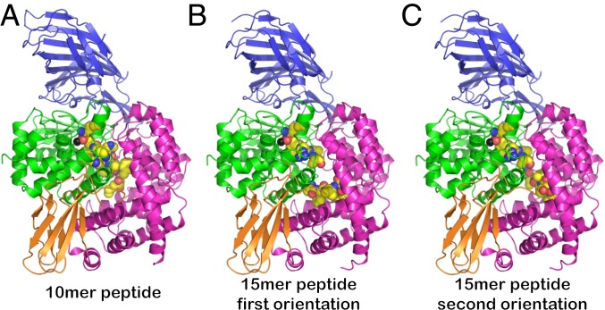
Fig. 1.
Cartoon representations of crystal structures of ERAP1 in complex with peptide analogs. The 4 domains of ERAP1 are shown in blue (domain I), green (domain II), orange (domain III), and magenta (domain IV). The catalytic Zn(II) atom is shown as a black sphere. The ligand is shown in spheres color-coded by atom type (yellow, carbon; blue, nitrogen; red, oxygen; orange, phosphorus). In all cases, the peptide is found trapped inside the internal cavity of the enzyme, with no apparent access to the outside solvent. (A) Crystal structure with the 10-mer peptide. (B and C) Crystal structures with the 15-mer peptide. The C-terminal moiety of the 15-mer was built in 2 distinct conformations. The middle part of the 15-mer peptide did not have a readily traceable electron density and is not shown here.

