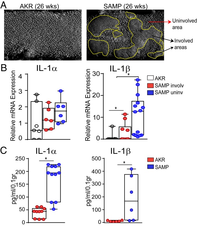
Fig. 1.
SAMP mice display increased expression of intestinal IL-1α and IL-1β. (A) Fixed ileal specimens analyzed by SM show cobblestoned abnormal mucosa in SAMP (Right) versus AKR (Left) mice. (B) IL-1α and IL-1β mRNA levels in healthy ileal mucosa of AKR mice and involved and uninvolved areas of SAMP mice. Data are presented as relative fold difference compared with AKR (arbitrarily set as 1). (C) IL-1α and IL-1β protein levels in tissue explants from ilea of SAMP versus AKR mice. Data are presented as median ± interquartile range. Statistical significance was determined by 1-way ANOVA followed by Dunn’s post hoc test, with n = 6–13, **P = 0.001 and *P = 0.016 (B); and by 2-tailed unpaired t test, with n = 6–10, *P < 0.01 (C).

