Fig. 3.
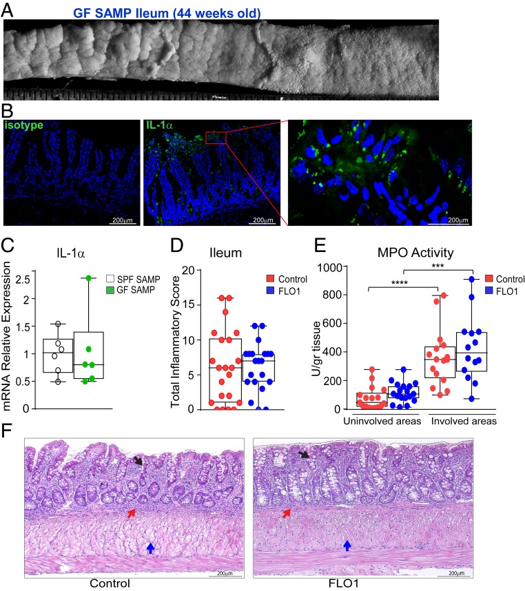
FLO1 treatment in GF SAMP mice has no detectable antiinflammatory effects. (A) Representative 3-D stereomicroscopy of cobblestoned mucosa in ileal tissues from GF SAMP, which, by immunofluorescence staining (B), show abundant nuclear-associated IL-1α protein (green) localized to surface IECs (n = 3). (Scale bars: Middle, 100 μm; Right, 25 μm.) DAPI is shown in blue. (Left) Isotype control staining. (Scale bar: 100 μm.) (C) IL-1α mRNA levels in ileal tissues from SPF and GF SAMP mice. FLO1- vs. vehicle (control)-treated SAMP mice evaluated by (D) total inflammatory score and (E) MPO activity in involved and uninvolved areas. (F) Representative photomicrographs of full-thickness H&E-stained ileal tissue from GF SAMP mice treated with either FLO1 (Right) or vehicle control (Left) show similar cellular infiltration (red arrows), mucosal thickening (blue arrows), and altered epithelial structure (black arrows). (Scale bar: 200 µm.) Data are presented as median ± interquartile range. In C, statistical significance was determined by 2-tailed unpaired t test (n = 6). In D–F, data are representative of 3 independent experiments and analyzed by 2-tailed unpaired t test (D) and Kruskal–Wallis followed by Dunn’s post hoc tests (E and F), n = 14–18; ****P < 0.0001 and ***P = 0.0005.

