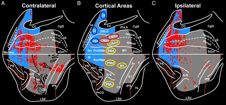
Fig. 2.
Origin of cortical inputs to the primate adrenal medulla. The survival time in this animal allowed retrograde transneuronal transport of rabies to label 6th-order neurons. The red squares indicate 200-μm bins with the highest numbers of labeled neurons (top 15%). (A) Flat map of the hemisphere contralateral to the injected adrenal medulla. The medial wall of the hemisphere is reflected upward and aligned on the midline. (B) Relevant areas of the cerebral cortex. Motor and somatosensory regions are shaded gray. Medial prefrontal regions are shaded blue. The cortical motor areas are indicated by yellow ellipses; cingulate motor areas that are involved in cognitive control are indicated by red ellipses; selected areas of the affective network in the medial prefrontal cortex are indicated by blue ellipses. (C) Flat map of the ipsilateral hemisphere. ArS, arcuate sulcus; CC, corpus callosum; CgG, cingulate gyrus; CgS, cingulate sulcus; CS, central sulcus; IPS, intraparietal sulcus; LSd, dorsal lip of the lateral sulcus; midline, junction between medial and lateral surfaces; mPF, medial prefrontal cortex; PrCO, precentral opercular cortex; PS, principal sulcus; RS, rostral sulcus; SGm, medial superior frontal gyrus; S1, primary somatosensory cortex. Numbers designate cytoarchitectonic regions. Adapted from ref. 11.

