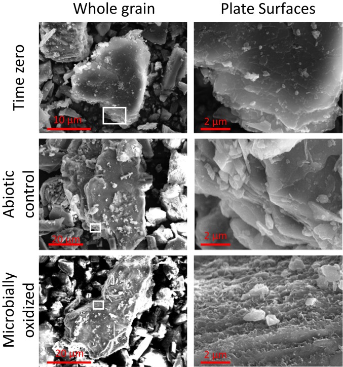
Fig. 4.
FE-SEM images of biotite at the whole-grain scale (Left) and basal plane (Right). Note the differences in scale on whole-grain images, as individual grain sizes are variable. For consistency, basal-plane images are at the same scale. The approximate area of the basal plane presented is outlined in white on the grain-scale images. Note the ragged appearance of the basal plane observed after microbial incubation.

