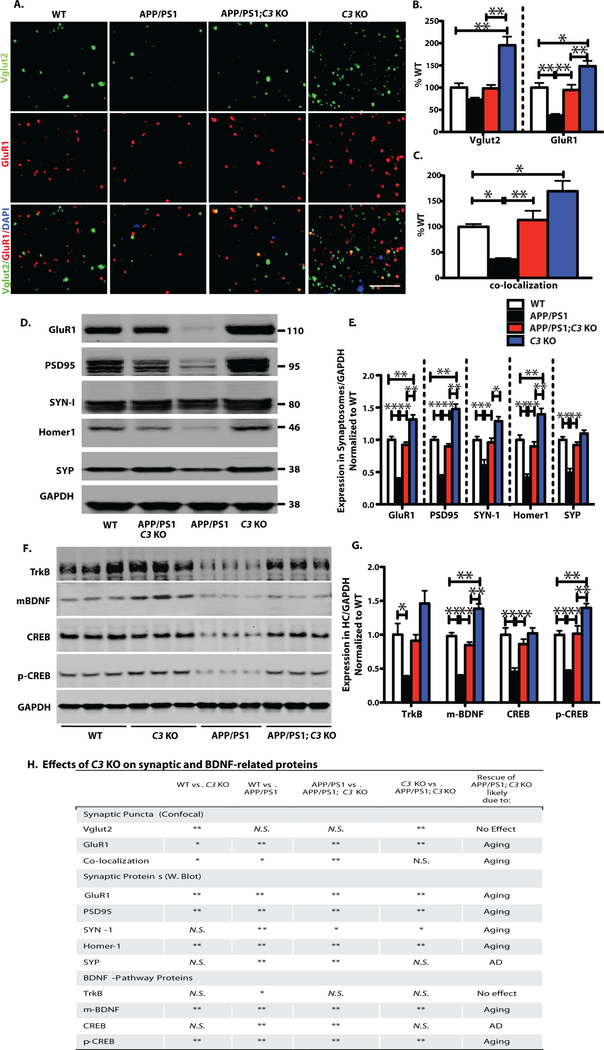
Fig. 5.
C3-deficiency resulted in partial preservation of synapse density and synaptic protein levels in APP/PS1 mice in spite of an increased plaque load. Comparisons were made between WT vs. C3 KO, WT vs. APP/PS1, APP/PS1 vs. APP/PS1;C3 KO, and C3 KO vs. APP/PS1;C3 KO (H). A. Synaptic puncta of pre-and post-synaptic markers Vglut2 and GluR1, respectively, and their colocalizaton in hippocampal CA3 were analyzed by high resolution confocal microscopy in 16-month-old mice. Scale bar = 5 μm. B. C3 KO mice had increased Vglut1 and GluR1 synaptic densities compared to WT, APP/PS1 and APP/PS1;C3 KO mice. APP/PS1 mice had significantly fewer GluR1 synaptic densities than WT mice while APP/PS1;C3 KO mice had significantly more GluR1 densities than APP/PS1 mice and were not significantly different than WT mice (* p < 0.05, ** p < 0.01; n = 6–8; 3 equidistant planes, 300 μm apart; one-way ANOVA and Bonferroni post-hoc test per marker). C. Colocalization of pre-and post-synaptic puncta reveals increased puncta in C3 KO vs. WT and APP/PS1 but not APP/PS1;C3 KO mice, reduced puncta in APP/PS1 mice vs. WT mice, and a rescue of synaptic puncta in APP/PS1;C3 KO mice compared to APP/PS1 mice, suggesting partial protection against synapse loss by C3-deletion (one-way ANOVA and Bonferroni post-hoc test). D,E. Western blotting of synaptic proteins in hippocampal synaptosomes isolated from aged mice indicates increased post-synaptic proteins GluR1, PSD95 and Homer 1 in C3 KO vs WT, APP/PS1 and APP/PS1;C3 KO mice. APP/PS1 mice had significantly lower post-synaptic GluR1, PSD95 and Homer1 and pre-synaptic SYN-1 and SYP compared to WT mice. APP/PS1;C3 KO mice had significantly more GluR1, PSD95, Homer1, SYN-1 and SYP than APP/PS1 mice, and were not significantly different than WT mice, suggesting a sparing of synaptic loss and normalization to WT levels in aged C3-deficient APP/PS1 mice (* p < 0.05; ** p < 0.01; n = 6; one-way ANOVA and Bonferroni post-hoc test per marker). F,G. Western blotting and quantification of TrkB, mBDNF, CREB, p-CREB in hippocampal homogenates of 16 month-old mice. C3 KO mice had increased mBDNF and p-CREB levels compared to WT, APP/PS1 and APP/PS1;C3 KO mice. APP/PS1 mice had significant reductions in all 4 markers compared to WT mice. APP/PS1;C3 KO mice had significantly higher mBDNF, CREB and pCREB than APP/PS1 mice, and all 4 markers were normalized to WT mouse levels (* p < 0.05, ** p < 0.01; n = 6; one-way ANOVA and Bonferroni post-hoc test per marker), suggesting a partial rescue of age-and/or AD-related lowering of BDNF pathway proteins. H. Table summarizing the effects of C3-deficiency on synaptic and BDNF-related proteins.

