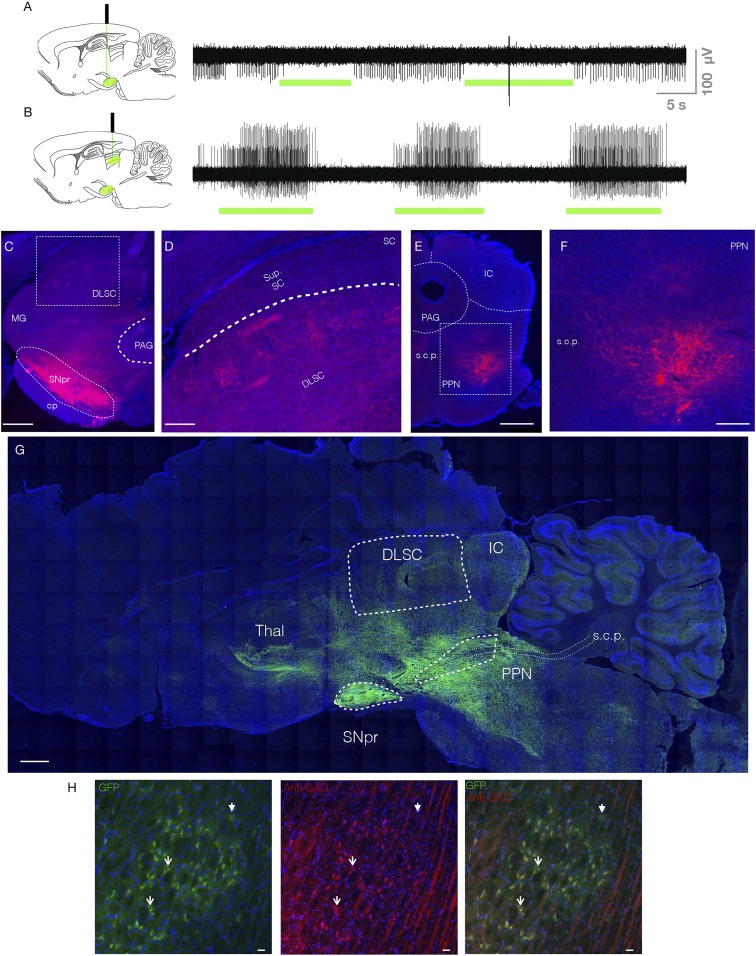
Fig. 1.
Electrophysiological and histological verification of opsins in the SNpr and its tectal and tegmental terminals. (A) Unit activity within SNpr. Green bars indicate optogenetic inhibition, which decreased in unit activity. (B) Unit activity within DLSC. Green bars indicate optogenetic inhibition of the nigrotectal terminals, which increased activity within DLSC. (C) Coronal section through SNpr and DLSC. Red indicates tdTomato fluorescence from the virus. Box indicates expanded view showing terminals in DLSC in D. (E) Coronal section through PPN. Box indicates expanded view showing terminals in F. (G) Parasagittal montage showing expression of GFP reporter in a GAD-Cre rat. Dense fluorescence is evident in SNpr, with fibers extending to known projections sites (DLSC, PPN, thalamus). Dotted lines outline structures of interest. (H) GFP+ cells (Left), anti-GAD immunofluorescence (Middle), and overlay (Right) showing that the majority of GFP+ cells colocalize with GAD. Solid arrowhead indicates a GAD−/GFP+ cell, open arrowheads indicate representative GAD+/GFP+ cells. PAG, periaqueductal gray; MG, medial geniculate nucleus; cp, cerebral peduncle; IC, inferior colliculus; Sup. SC, superficial layers of SC; s.c.p., superior cerebellar peduncle; PPN, pedunculopontine nucleus; Thal, thalamus. (Scale bars: C, E, and G, 1 mm; F and D, 330 μm; H, 25 μm.)

