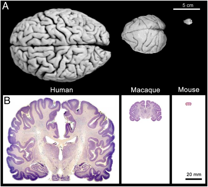
Fig. 1.
Comparison of human, macaque, and mouse brains. (A) Images of the dorsal surface of a human brain, macaque brain, and mouse brain. Notice the striking differences in the size and convolution complexity of the cerebral cortex across the 3 species. (B) Coronal Nissl-stained sections of hippocampus-containing tissue in the 3 species. Reproduced with permission from ref. 104.

