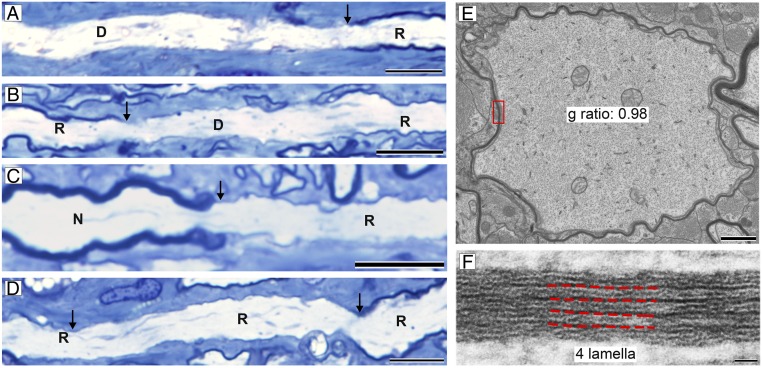
Fig. 6.
Short internodes with thin myelin in partially recovered ONs. (A–D) Longitudinal sections at level 1 showed that many internodes were short and thin, demyelinated (D), remyelinated (R), or normal (N) (arrows mark nodes or heminodes). (E) EM examination of the ON at level 2 showed that many thinly myelinated axons had 5 or fewer myelin lamellae. (F) Boxed area from E. (Scale bars: A–D, 20 µm; E, 1 µm; and F, 20 nm.)

