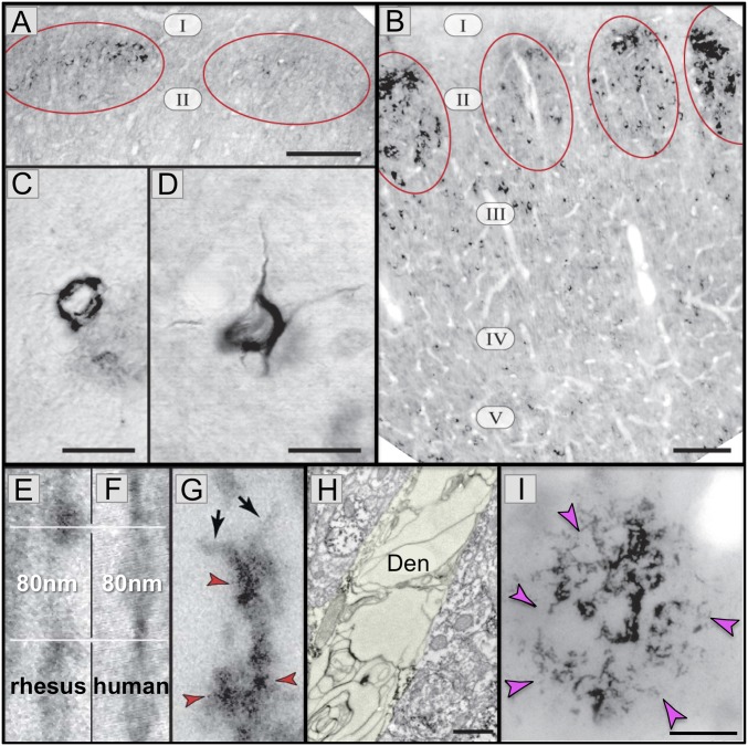
Fig. 2.
AT8 pTau and amyloid pathology in aged rhesus monkeys (26 to 38 y). (A) AT8 label is first seen in layer II cell islands in the ERC in a 26-y-old monkey. (B) In an older monkey (33 y), AT8 labeling is more extensive in layer II and now extends to deeper layers. (C and D) Higher-magnification view of neurofibrillary tangles in layer II (C) and layer V (D) from a 38-y-old monkey. (E–G) High-magnification view of AT8-labeled (red arrrowheads) paired helical filaments from an aged monkey (E and G) or human (F). Note the same width (10 nm) and helical frequency (80 nm). Black arrows in G show the abrupt ends of each filament. (H) A degenerating dendrite (Den) from an AT8-labeled neuron in aged monkey layer II ERC. The dendrite is devoid of normal organelles and is filled with autophagic vacuoles similar to those in LOAD. (I) An Aβ-labeled (magenta arrowheads) amyloid plaque from layer V ERC in an aged monkey. Reprinted from ref. 7, with permission from Elsevier. (Scale bars: A and B, 100 μm; C and D, 10 μm; E–G, 40 nm; H, 500 nm; I, 20 μm.)

