Fig. 7.
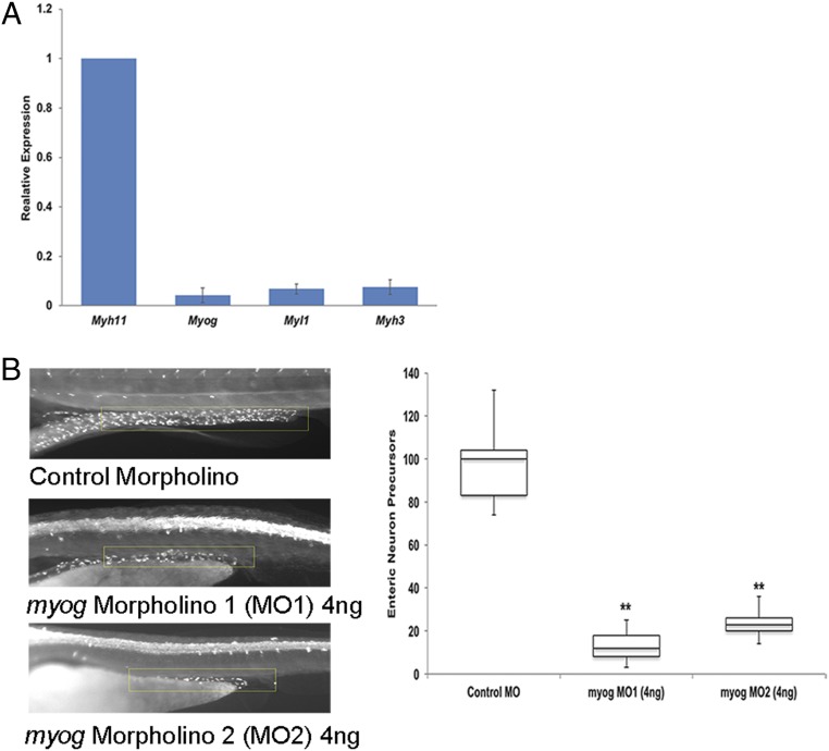
Crosstalk between enteric neurons and their supporting mesenchymal cells. (A) Gene expression qPCR of specific muscle markers was performed using FACS-sorted Mhy11-positive cells from Myh11-EGFP transgenic mouse guts at E10.5. Low-level expression of Myog, Myl1, and Myh3 is observed in the gut smooth muscle cells at this stage of development. (B) Morpholino knockdown of myog in zebrafish leads to a skeletal muscle phenotype (significant body curvature along the anterior–posterior axis). Morpholino knockdown of myog in zebrafish leads to the skeletal muscle phenotype of significant body curvature along the anterior–posterior axis along with significant loss of enteric neuron precursors at 4 dpf. The neuronal numbers, counted in a region of interest starting at the eighth somite and measuring until the end of the gut tube (yellow box), upon sampling 50 embryos each from wild-type and morphant embryos are significantly different (**P < 0.001).

