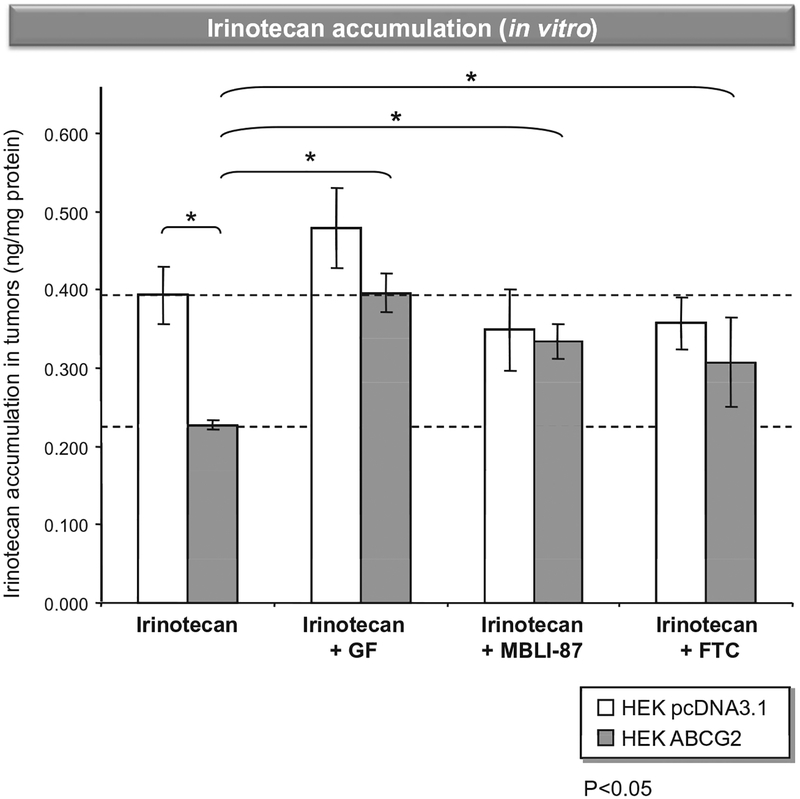
Fig. 1 –
In vitro irinotecan accumulation in control and ABCG2-expressing cells. Cells were loaded with 2 μM irinotecan in DMEM medium (without FBS) for 60 min at 37 °C in the absence or presence of either 5 μFM GF120418, 5 μM MBLI-87 or 10 μM fumitremorgin C. Intracellular irinotecan accumulation was quantified by HPLC MS/MS. Results were expressed as ng irinotecan/mg protein.

