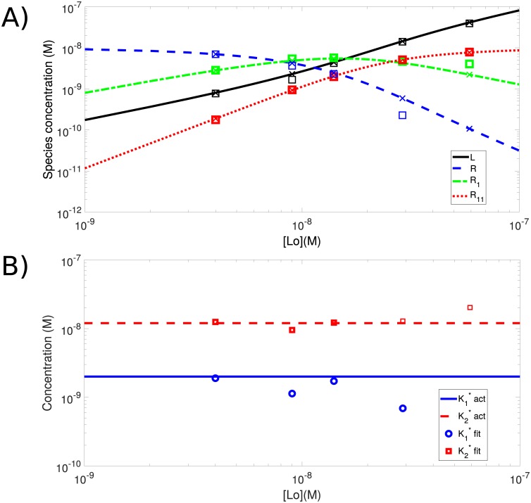
Fig 6. Spatial cPCH can be used to estimate the dissociation constants in a receptor-ligand system with two different binding sites on the receptor and a fluorescent ligand.
A) Spatial cPCH can accurately estimate concentrations if the concentration of the doubly-labeled receptor R11 is lower than that of the singly-bound receptor R1. The actual concentrations are indicated with crosses and the estimated concentrations are indicated by the open squares. The initial concentration of receptors is R0 = 10 nM. We specified the (apparent) dissociation constants for the singly and doubly bound receptors as and and find the actual dissociation constants from Eq S9. These apparent dissociation constants arise because we cannot distinguish using either fluorescence or diffusion which site the ligand has bound on a receptor that has bound only one ligand (Sec. S5). The simulation employed a laser dwell time of 10 μs with images of 512 × 512 pixels and an image size of 4 μm by 4 μm, so that the pixel size is just under 12 nm. The line retracing time is 1 ms and the frame retracing time is 50 ms. We generated 2 sets of data with 25 images each for all of the concentrations used, except for the lowest initial value of ligand, which used 1 set of 70 images. B) The apparent dissociation constants ( and ) can be estimated using the measured concentrations provided the concentration of singly labeled receptors R1 is greater than the concentration of doubly labeled receptors R11.

