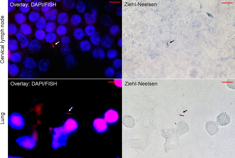
Fig 2. Microscopic images of imprint slides prepared from lung and cervical lymph nodes of Mycobacterium tuberculosis-infected mice at day 28 post-infection with combined FISH, DAPI and Ziehl-Neelsen staining.
For fluorescent in situ hybridization (FISH), the slides were observed using a red channel of a Leica DMI6000 microscope under a 100 X oil-immersion objective. The images were captured in the same microscopic field using a Hamamatsu Orca AG camera (Hamamatsu Photonics, Herrsching-am-Ammersee, Germany) for FISH-positive mycobacteria (left images, white arrows) and a DFC425 C Digital Microscope Camera (Leica Microsystemes, Nanterre, France) for Ziehl-Neelsen-positive mycobacteria (right images, black arrows). Scale bar = 5 μm.

