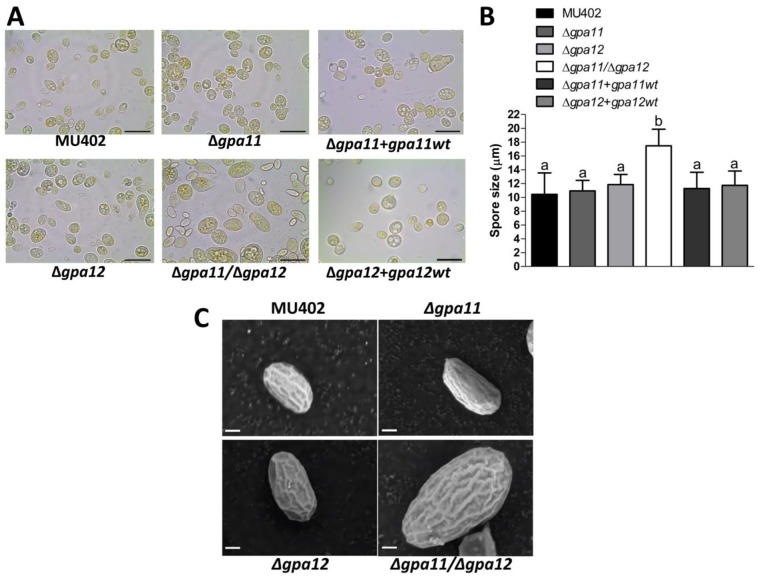
Fig 4. Influence of gpa11 and gpa12 on sporangiospore size in M. circinelloides.
The spores from the different M. circinelloides knockout strains produced in YPG were observed under A) light microscope (100 X), scale bar equal to 20 μm. B) The spore size of each strain was quantified using the Leica Application Suite. C) The spores obtained after five days of incubation on solid YPG media were observed under scanning electron microscope. Representative photographs from the corresponding strains of M. circinelloides under 10,000 magnifications. Scale bar is equal to 1 μm. Statistically significant differences are indicated by different letters (ANOVA and Fisher’s tests; p ≤ 0.05).

