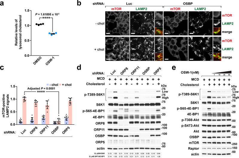
Fig. 2 |. OSBP is required for cholesterol-dependent lysosomal recruitment and activation of mTORC1.
a, OSBP inhibition by OSW-1 treatment reduces the lysosomal cholesterol content. HEK-293T cells expressing Tmem192-mRFP-3xHA were treated with DMSO or 20 nM OSW-1 for 8h. Lysosomes were purified and analyzed by mass spectrometry (mean ± s.d., n = 4 biologically independent samples per treatment, two-tailed, unpaired t-test. ****P = 1.61895 × 10⁻⁶ vs. DMSO). See immunoblots in Supplementary Fig. 4a. b, OSBP is specifically required for lysosomal recruitment of mTORC1 by cholesterol. Cells depleted of OSBP were subjected to cholesterol depletion and restimulation, where indicated, followed by immunofluorescence for endogenous mTOR and LAMP2. Representative images are shown. Scale bar, 10 μm. c, Quantitation of co-localization of mTOR with LAMP2-positive lysosomes in cells expressing the indicated shRNAs (mean ± s.d., 10 fields of view per genotype/condition; n represents cell number: shLuc – chol (n = 182), shLuc + chol (n = 181), shORP8 – chol (n = 104), shORP8 + chol (n = 91), shORP11 – chol (n = 157), shORP11 + chol (n = 156), shOSBP – chol (n = 176), shOSBP + chol (n = 182), shORP5 – chol (n = 160), shORP5 + chol (n = 166), ANOVA with Dunnett’s multiple comparison test. ****Adjusted P = 0.0001 vs. shLuc + chol). See representative images in Supplementary Fig. 6a. d, ORPs affect mTORC1 activation by cholesterol. Cells were sterol-depleted using methyl-beta cyclodextrin (MCD, 0.5% w/v) for 2h and restimulated for 2h with 50 μM cholesterol complexed with 0.1% MCD (MCD:cholesterol). Cell lysates were analyzed for the levels of indicated proteins and for phosphorylation status of S6K1 (T389), 4E-BP1 (S65). Fold changes of protein phosphorylation are indicated. e, OSW-1 inhibits sterol-induced mTORC1 signaling in a dose-dependent manner. Cells were subjected to cholesterol starvation and restimulation in the presence of DMSO or OSW-1 at the indicated concentrations and immunoblotted for the indicated proteins. Experiments in b, d performed three times and in a, e two times. See unprocessed blots in Supplementary Figure 9. Statistics source data are provided in Supplementary Table 2.

