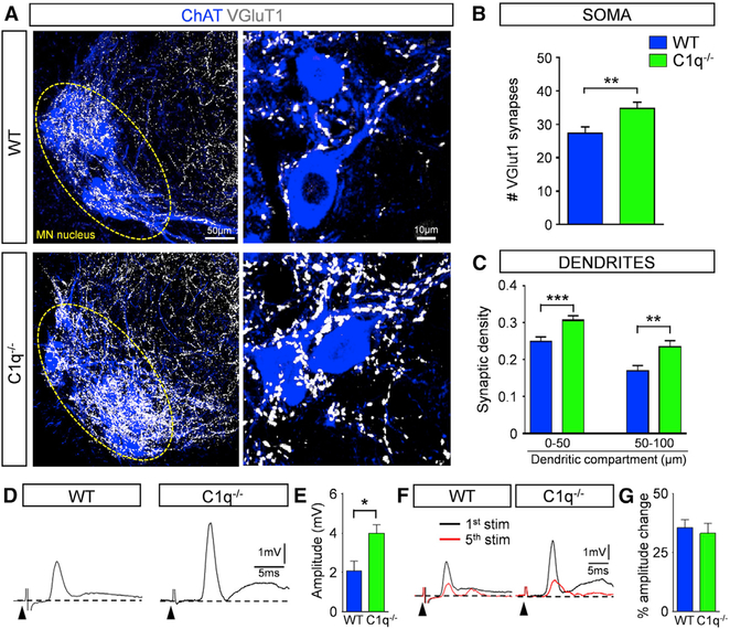
Figure 6. Genetic Deletion of C1q Results in an Increase in Proprioceptive Synapses during Normal Development.
(A) z stack projections of confocal images of ChAT (blue) and VGluT1 synapses (white) in WT and C1q−/− mice at P11 (total distance in z axis: 25.2 μm). Circled areas indicate MN nuclei. Higher magnification (right) of z stack projections from single MNs in WT and C1q−/− mice (total distance in z axis: 2.1 μm).
(B) The total number of VGluT1 synapses on the entire soma from WT (n = 10) and C1q−/− (n = 12) L1 MNs. **p < 0.01; t test.
(C) VGluT1 synaptic density (number of synapses in 50-μm dendritic compartments from the soma) in WT (n = 41) and C1q−/− (n = 35) MNs. **p < 0.01, ***p < 0.001; t test.
(D) Ventral root responses to supramaximal stimulation of the homonymous L1 dorsal root in WT and C1q−/− spinal cords at P10. The arrowheads indicate the stimulus artifacts.
(E) The amplitude of the monosynaptic reflex responses for WT (n = 6) and C1q−/− (n = 7) mice at P11. *p < 0.05; t test.
(F) Superimposed traces of the 1st (black) and 5th (red) responses following 20-Hz stimulation of the dorsal root in WT and C1q−/− mice.
(G) Percentage amplitude change of the 5th response normalized to the 1st response, following 20-Hz stimulation in WT (n = 6) and C1q−/− (n = 7) mice.

