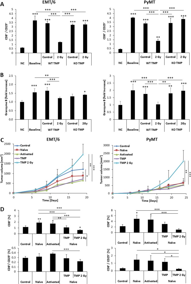
Fig. 2.
TMPs shed from irradiated EMT/6 and PyMT cells inhibit cytotoxic T-cell activity. a Splenocytes isolated from naïve 8–10-week-old BALB/c mice were cultured with CD3+/CD28+ T-cell activating beads in the absence (baseline) or presence of TMPs derived from the control or irradiated WT EMT/6 or PyMT cells or their PD-L1 KO counterparts as indicated. Negative control (NC) refers to cultures in the absence of activation beads. Twenty-four hours later, the splenocytes were harvested and the percentage of activated cytotoxic T cells (CD8+/CD25+) was evaluated by flow cytometry. b Granzyme B levels in the conditioned medium of the cultures described in (a) were assessed by specific ELISA. The results represent three biological repeats. c, d Eight to 10-week-old SCID and 6 Gy sub-lethally irradiated C57Bl/6 mice (n = 5 mice/group) were implanted with EMT/6 or PyMT cells mixed with splenocytes from untreated (naive) mice in a 1:100 ratio. In addition, in some groups, these samples were mixed with TMPs originated from control or 2 Gy irradiated cells, and subsequently injected into mice. A mixture of EMT/6 or PyMT cells with activated splenocytes from tumor-bearing mice were used as a positive control. EMT/6 and PyMT implanted to the mammary fat pad alone were used as negative control. Tumor growth was assessed twice weekly (c). At end point, tumors were extracted and the percentage of the total and activated CD8 cells (CD8+/CD25+) was evaluated by flow cytometry (d). *p < 0.05, **p < 0.01, ***p < 0.001, as assessed by one-way ANOVA followed by Tukey post hoc test

