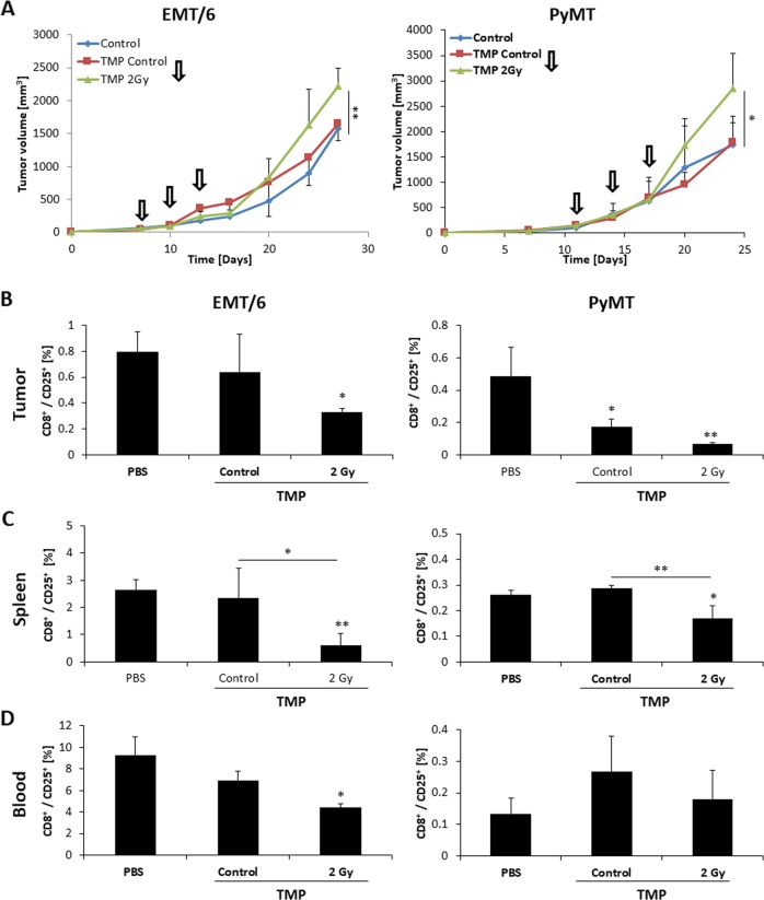
Fig. 3.
TMPs from irradiated EMT/6 or PyMT cells promote tumor growth and inhibit cytotoxic T-cell activity in vivo. a EMT/6 or PyMT tumor cells (5 × 10⁵/mouse) were orthotopically implanted into the mammary fat pad of 8–10-week-old female BALB/c and C57Bl/6 mice, respectively (n = 4–5 mice/group). When tumors reached ~100 mm3, mice were either intravenously injected with PBS (control) or TMPs derived from control or 2 Gy irradiated EMT/6 or PyMT cells every 3 days (indicated by arrows, 1 × 10⁵ TMPs/mouse). Tumor volume was monitored twice weekly. b–d At end point, blood was drawn, and tumors and spleens were harvested. The percentage of activated CD8 cells (CD8+/CD25+) was evaluated in tumors (b), spleens (c), and peripheral blood (d) by flow cytometry. *p < 0.05, **p < 0.01, as assessed by one-way ANOVA followed by Tukey post hoc test

