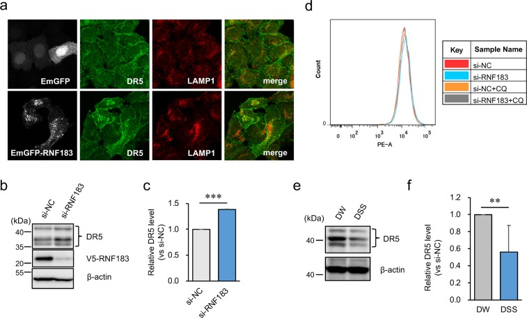
Figure 5.
Involvement of RNF183 in the localization and degradation of DR5. (a) Effect of RNF183 overexpression on DR5 subcellular localization. HeLa cells transfected with EmGFP-tagged RNF183 or EmGFP alone were subjected to immunofluorescence staining with DR5 and LAMP1 antibodies (grey, EmGFP; green, DR5; red, LAMP1). (b) Effect of RNF183 knockdown on DR5 protein levels. RNF183-siRNA or non-target control (NC)-siRNA were transfected into HEK293 cells stably expressing V5-tagged RNF183. Cell lysates were subjected to western blotting with DR5 and V5 antibodies. The full-length blots are presented in Supplementary Fig. S10. (c) Quantitative graph of data from (b). Asterisks represent significant differences (n = 3; paired t-test, ***p < 0.001). (d) Cell surface expression level of DR5. HEK293 cells expressing V5-tagged RNF183 were treated with 100 μM CQ for 12 h. Then, cells were labeled with PE-conjugated anti-DR5 antibody. (e) DR5 protein levels in colonic epithelial cells from DW- and DSS-treated mice at 5 days. Cell lysates were subjected to western blotting with anti-DR5 and anti-β-actin antibodies. The full-length blots are presented in Supplementary Fig. S11. (f) Quantitative graph of data from (e). Asterisks represent significant differences (n = 6; paired t-test, **p < 0.01).

